Ultrasound examinations during pregnancy
Ultrasound examination is safe for the expectant mother and the child. During the examination, the child is not stressed. Until now, no confirmation of harmful effects of USG examinations have been received from patients or specialists that perform such examinations on daily basis. The current data confirm that reasonable use of USG has considerably more benefits than potential side effects, if any. USG can be performed as many times as you would like, but you have to clearly understand the objectives and purpose of each examination, and any and all USG examinations should have clear medical indications. To prepare for the USG examination, the patient does not have to take any particular steps. However, to assess the cervical canal better it is advised not urinate before the examination.
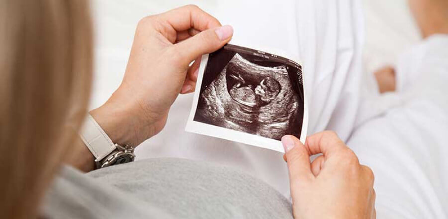
During the pregnancy, women often feel fear and anxiety about the health of the unborn child, and worry about correct development of the foetus. To make sure that the child is fine, ultrasound screening examinations are needed.
During the pregnancy, ultrasound examinations allow to:
- Exclude the most important and dangerous foetal development pathologies
- Find a failed pregnancy
- Accurately determine the onset of pregnancy in case of irregular menses or when the time of conception is unknown
- Timely diagnose pregnancy complications
- Control foetal development and wellbeing
How often should ultrasound (USG) examinations be performed during the pregnancy?
During a pregnancy, three ultrasound examinations are required:
- Week 11 to 13 of the pregnancy (I trimester)
- Week 20 to 22 of the pregnancy (II trimester)
- Week 32 to 34 of the pregnancy (III trimester)
USG diagnostics is particularly important during I trimester (Week 11 to 13). During this developmental stage, USG already allows to see signs of congenital developmental pathologies (incl. chromosomal pathologies, cardiac diseases, etc.)
USG examination before a delivery is required very rarely. It includes cases when the acquired information may affect the delivery management tactics, usually when the course of the pregnancy is difficult. Indications are determined by the responsible doctor.
Pre-natal diagnostics that is based on USG examination focusses on early exclusion of severe congenital anomalies. If a pathology is found during I trimester, the patient can terminate the pregnancy during an early foetal development stage that is less harmful for her health. If the woman chooses to keep her pregnancy, she and her partner has enough time to prepare for the birth of a child with special needs that will require specialized care since the first days of its life. The doctors inform about early rehabilitation opportunities for children with congenital anomalies, as well as the risks and consequences of the parental decision.
Pregnancy ultrasound — new US diagnostics opportunities at iVF Riga: the expert ultrasound system Voluson E8
To follow the latest developments, we keep upgrading our equipment, and offer ultrasound examinations during pregnancy with the last generation Voluson E8 system: a very sensitive system that supports a wide range of examinations.
Advantages of the Voluson E8 ultrasound system:
- Quick processing of data, clear and detailed image visualization
- High contrast and resolution in combination with various scanning regimes
- Excellent visualisation quality in any mode: from 2D images to the latest 3D/4D technologies
- Extensive scanning area to see the smallest of details
- Sensitive colour doppler to study circulation in vessels during echocardiography
- Improved HDlive technology that allows to combine several independent light sources and produce hues and shades for high-precision diagnostics that allows to generate anatomically accurate images
- Innovative imaging mode for vessel structures
- Ability to create reconstructions of surface vessels, as well as to evaluate the vessel wall structure from inside to gain a deeper insight into the topographic anatomy of the circulation system and the surrounding structures
- Technologies that help in performing semi-automatic, standardized foetal neck and nuchal fold thickness measurements during I trimester by reducing manual measurement errors that is particularly important during high-risk pregnancies
- Ability to generate reports and record studies in real time, incl. on USB drives; ability to e-mail ultrasound data in the safe mode
After the long-awaited pregnancy, foetal development is very important. It requires specialist monitoring, as well as specialized examinations at various gestational stages. To monitor pregnant patients, it is very important to choose experienced and skilled specialists who can operate the most advanced diagnostic devices to monitor foetal development and condition, notice early signs of development and recognize even the slightest deviations from the normal.
Doctor of Medical Sciences, Associate Professor of Clinical Medicine, Obstetrics and Gynaecology; experienced ultrasonography specialist Dr. Natālija Vedmedovska performs pregnancy ultrasound diagnostics at our clinic.
 en
en lv
lv ru
ru lt
lt se
se no
no

